Animal Cell
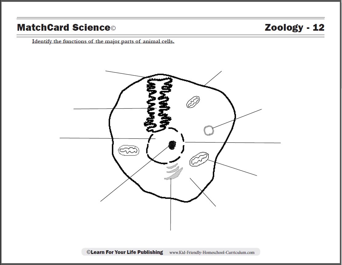
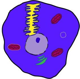
The animal cell diagram identifies functions of the major parts of the cell.
Free Download Below
Animal Cell MatchCard
Teach the 10 Parts of Animal Cells
Objective: Identify the functions of ten major parts of animal cells.MatchCard: Download below.
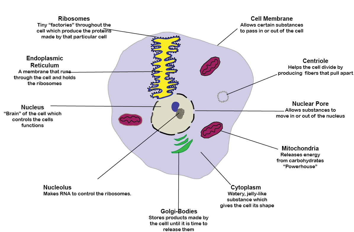
MatchCard Information Pieces identify ten cellular parts and their function which are matched to the animal cell diagram.
Print the Animal Cell MatchCard

Click image to go to download.
This is MatchCard #12 of the Zoology Unit Study. Find more information on MatchCard Science below.
Introduce Animal Cells
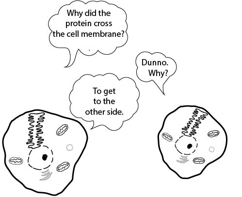
It's A 3-D World: Give Your Students the Artistic Challenge
Show the animal cell diagram to the student(s) and ask what they think it is. Some students may be able to identify some of the structures.An orange makes a great illustration of the three dimensional nature of cells. Cut the orange in half. Have the students draw a diagram of the cross-section of the orange.
Compare the cross-section of the orange diagram and the cross-section of the animal cell diagram. Can they explain where the term "cross-section" comes from?
Organelles on the Animal Cell Diagram
Day 1: Cell Membrane, Nucleus, Cytoplasm
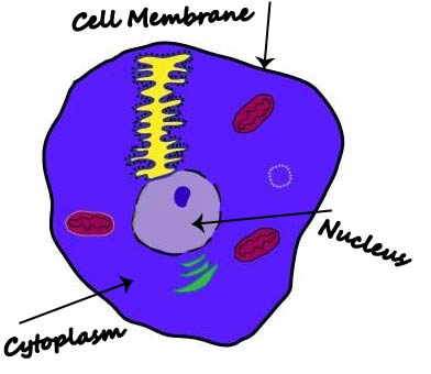 The first three parts of the cell to explain are the cell membrane, nucleus, and cytoplasm.
Cell Membrane
The cell membrane is the "shell" of the cell. We can compare the orange's peel to the cell wall of a plant (animals don't have the cell walls.) The white membrane inside the peel and surrounding the fruit can be compared to a cell membrane - which are found on both plant cells and animal cells.
Nucleus
The nucleus is the "brain" of the cell. It is the headquarters that controls all the functions. To some extent it might be compared to the central fibrous section of an orange; but it's function is closer to the brain of an animal.
Cytoplasm
The cytoplasm is the "jelly" that gives the cell is shape. It can be compared to the juice in the orange.
Hands-On Projects
Demonstrate Cell Membranes with Orange Membranes
Take one of the sections of an orange and cut it in half. Try to scoop as much of the fiber and juice out of the "bag". Discuss how substances can go through the membranes.Demonstrate Cytoplasm with Gel
Demonstrate the cytoplasm of a cell with the toy gel balls you can get in many toy stores. You can also squeeze the small ketchup containers from fast food restaurants to try and imagine the gel inside of a cell.This is the first lesson on the animal cell. For kids in 3rd and 4th grade, this may be all the information they need. For students in fifth grade and above, continue the other lessons in a separate session.
Day 2: Mitochondria, Golgi Bodies, Nuclear Pore, Centriole
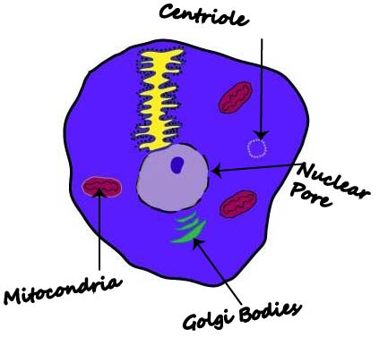 The second lesson introduces students to four distinct organelles of the cell.
Mitochondria
The mitochondria are considered the "powerhouse" of the cell. They convert carbohydrates to energy the cell can use.
Students who have done the Botany Unit Study may be reminded of how starch is used in the carbon cycle. The starch (or carbohydrates) is converted inside the mitochondria of the cells.
Golgi Bodies
The cell is like a miniature factory or city. The golgi bodies store the different chemicals made by different parts of the cells until they are ready to be released into the cytoplasm. The golgi bodies are fairly easy to see under the microscope of many types of cells.
Nuclear Pore
In the last lesson you discussed cell membranes on the outside of the cell and the nucleus in the inside. There is also a thin nuclear membrane surrounding the nucleus. It has large pores which can be compared to the pores of swiss cheese. These pores allow substances to leave the nucleus.
Centrioles
The centrioles are stringy fibers which are used in cell division. The fibers help to pull the two parts of a cell apart when it is dividing.
Day 3: Endoplasmic Reticulum, Ribosomes, Nucleolus
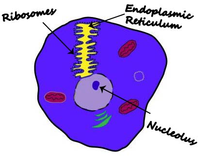 Middle School students can continue their study of animal cells by learning about the ribosomes which make proteins for the cell.
Every type of cell is different. Eye cells produce tears, pancreatic cells produce insulin, stomach cells produce acids. The types of protein the cells make will depend on the cell itself. However, the basic cell parts are similar.
Ribosomes
The Ribosomes are small "factories" that produce the proteins made by that cell. Proteins can be compared to words made from letters. 26 letters of the alphabet make an infinite number of cells. Four nucleotides make an infinite number of different proteins. This happens in the small ribosomes inside the cell.
Endoplasmic Reticulum
The endoplasmic reticulum is a membrane that is folded back and forth inside the cell but outside of the nucleus. It can be compared to the mesh inside an orange or grapefruit. The ribosomes are lined up along the endoplasmic reticulum.
Nucleolus
The nucleolus is an organ within the nucleus. It makes a substance call ribosomal RNA. This controls how the ribosomes make proteins.
Make a 3-D Model of a Cell
A great way to learn about cells is to make a model of one. You can buy kits to make cell models.You can also make a clay animal cell model. Use different colors of clay for the different cellular structures.
Your artistic students might also like to diagram their own 3 dimensional cell using colored pencils or other media.
MatchCard Science
How To Use MatchCards
Download the FREE MatchCard Science Instructor's Guide and see how MatchCards can make building their science knowledge base fun.
12 Science Unit Studies

Chemistry is only one of twelve complete unit studies for kids in 3rd to 8th grade.
Comprehensive objectives, hands-on projects, suggested science fair experiments, and the fun game-like MatchCards keep them interested in learning science. See all twelve MatchCard Science Unit Studies.
About Our Site
Hands-On Learning
