Circulatory System
Circulatory System Lessons for 3rd - 8th Grade
Our Heart and Lung Lesson Plans includes the MatchCard Worksheet and hands on projects.
Free Download Below
Circulatory System for Kids
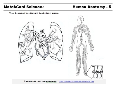
Objective: Trace the route of blood through the circulatory system.The MatchCard consists of a Circulatory System Diagram. The students will trace the flow of oxygenated and unoxygenated blood through the heart, lung, and body.
Download and Use the Circulatory System MatchCard
This is MatchCard #5 of the Human Anatomy Unit Study. Directions for using MatchCards are below.Introducing the Circulatory System for Kids
The Analogy of An Inflatable Pump
For starters, take a sheet of paper and fold it in half one way, and half the other way, to make four quarters (two on top and two on bottom.) You will use this is in a few minutes.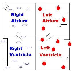
Have you ever been in an inflatable room, the type you see at carnivals that children can jump in? Let's imagine that the human heart is like a giant, inflatable house.
Our house is going to have two stories, with two rooms on the top, and two rooms on the bottom. The rooms are side by side. (Show paper you folded.) There will also be doors, and windows, and chimneys in our house.
Our house is inflatable, so it can squeeze and get larger and smaller. That is what the real heart does.
The heart sends blood through the whole body, and the blood contains red blood cells which carry oxygen.
With the analogy of our inflatable house, we will imagine that little empty balloons go into the house without oxygen, and round ballons filled with oxygen leave the house.
We are going to talk about the four rooms in the heart and how they work together to get blood to the whole body. We are going to start by talking about the rooms on the right.
Write "Right" on both the right upper and right lower boxes. This will be on the left hand side of the boxes you are working with. See the information in the box below if you need to clarify the right-left orientation.
Right and Left Sides
If you are ever looking at a diagram of the human body, you may notice that it says "right" on the left side of the page; and "left" on the right side of the page.There's a simple reason for it. The diagram is a diagram of another person. If you are standing and facing someone, their right hand is on the opposite side of your right hand.
Imagine you are in the inflatable house. There are rooms on the right side.
However, if you go out of the house and turn around and face it, those rooms are now on your left.
Whenever you see an anatomical drawing, it is as if you are facing that person, so their right and left are opposite of yours.
In the right upper room, there is a window at the top. Lots of empty balloons fall into this room.
Draw flat ovals in the right upper box to represent empty balloons - or unoxygenated red blood cells.
There is a trap door between the upper and lower rooms in the right heart. When the upper rooms are squeezed, this trap door opens, and all the empty ballons fall into the right lower room.
When the upper rooms are no longer being squeezed, the bottom rooms get squeezed. When this happens, the empty balloons in the bottom of the right heart get pumped up the chimney, and out of the house. Those balloons go through some tunnels to air tanks near the lungs.
Once the balloons are filled with air, they come back to the house. But this time, they come in through a back door in the upper room of the left heart.
Draw a small door, marked "back door" in the upper room of the left heart, and show round little balloons in this room.
When the right upper heart gets squeezed, the left upper heart gets squeezed also. At that time, the trap door between the upper and lower rooms opens, and the filled balloons fall into the bottom room of the left heart.
Draw trapped door, and filled balloons on the left side.
Finally, when the bottom of the right heart is squeezed; the bottom of the left heart is squeezed also. Then, the balloons in the bottom left room go up a chimney are sent out of the house, and into the whole neighborhood.
In this case, the neighborhood is the rest of the body. The job of the red blood cells is to take oxygen to every cell in the body.
Circulatory System Worksheet
Show the Circulatory System MatchCard and the diagram of the heart and lungs. Compare it to the simpler boxed diagram.The important point is to recognize unoxygenated blood comes to the right side of the heart, goes to the two lungs on either side for oxygen, and then returns to the left side of the heart.
In the diagram of the heart, you will see flat lines to represent the unoxygenated blood, and small circles to represent oxygenated blood. Traditionally, blue is used to color areas where there is unoxygenated blood, and red for the areas of oxygenated blood. Students may color their MatchCards to help them grasp the difference. Red and blue color pencils that have been sharpened will work best.
Lungs
The lungs are the "air stations" on either side of the heart. As you take a deep breath, oxygen goes into the respiratory system. That oxygen moves across the cell membranes into the blood that will return to the heart.Encourage the students to color the blood vessels going from the heart to the lungs blue; and the oxygenated blood returning to the heart red.
Cut apart the first four information pieces, and have the students identify the first four steps in the circulation:
- Right side of heart
- Lungs
- Left side of heart
- Aorta
Arteries
Blood vessels are like small hoses in the body. The arteries are the blood vessels that take the oxygenated blood that has left the heart and distributes it to the rest of the body.On the circulatory system diagram, two types of vessels can be seen: arteries and veins. The arteries are drawn with black lines, and the veins with lighter, gray lines.
Color the arteries through the body red.
Branching blood vessels
Arteries branch into smaller and smaller vessels. Small arteries are called "arterioles."The branching system of vessels can be compared to tree trunks, large branches, small branches, and twigs. The concept is that arteries move from large (the heart) to small (the cells of the body.)
Capillaries and Cells
The very smallest blood vessels are the capillaries. These thread like vessels are so tiny they only allow one red blood cell to move from an arteriole into a cell. Every cell in the body has capillaries to take oxygenated red blood cells and other nutrients to it.The Venous System
The veins are the opposite of the arteries. They carry the red blood cells away from the cells. Since the cell "took" the oxygen, they are unoxygenated.When the blood leaves the cells, it goes into through the capillaries into the very tiny venules. Then it moves to small veins, to larger veins, and finally through the large vena cava back into the heart.
Color the veins blue in the diagram on the MatchCard.
Circulation
The steps of circulation once blood has left the heart is:- Arteries
- Arterioles
- Capillaries
- Venules
- Veins
- Back to the heart
Cut the other MatchCard Information pieces, and match them to the diagrams.
Activities on the Circulatory System For Kids
Take Your Pulse
When the heart beats, the chambers become smaller to squeeze blood through the valves, which are like trap doors between the rooms.You can feel your pulse at different parts of the body. The easiest to find is on your wrist on the same side as your thumb.
Find your pulse and count it for 60 seconds.
Then go and run around your house five times. If it is too cold out, you might try one of these other exercises:
- Run up and down the stairs three times
- Do 30 jumping jacks
- 20 push ups
It is both faster and stronger.
Did you also notice you were breathing faster and heavier? Why?
Listen to the Beats
If you can, get an inexpensive stethoscope to listen to your own heart beat.You will hear two sounds instead of one when the heart beats. Sometimes people say it sounds like the heart is saying "lub dub". What do you think it sounds like?
However, if you take your own pulse, you only feel one beat.
The two sounds are caused by the valves, or trap doors, in the heart. The first sound is the valves between the upper and lower parts closing. That prevents blood that is the ventricles (bottom rooms) from moving back up into the atrium. This is the "lub" sound.
The second sound is the valves that prevent blood that has just left the heart through the ventricles from going back into the ventricles. That is the "dub" sound.
Cook Up Some Blood
Yuck! Sounds gross!Actually, this won't be gross at all. You will need:
- Red gelatin mix
- Red M&M's or other candy (represent red blood cells)
- Small white marshmellows (represent white blood cells)
- Sesame seeds (represent platelets)
Here are the directions:
- Make the gelatin according to the directions on the box.
- When it is time to add other ingredients, add the three items listed above: M&M's, marshmellows, sesame seeds
- Put in the fridge to thicken
- Red Blood Cells - carry oxygen to every cell in your body
- White Blood Cells - Fight diseases, including bacteria and viruses
- Platelets - Make a sticky substance that turns into a scab so you don't loose all your blood when you bleed.
MatchCard Science
How To Use MatchCards
Download the FREE MatchCard Science Instructor's Guide and see how MatchCards can make building their science knowledge base fun.
Human Anatomy Unit Study
Download the entire Human Anatomy Unit Study
12 Science Unit Studies

Chemistry is only one of twelve complete unit studies for kids in 3rd to 8th grade.
Comprehensive objectives, hands-on projects, suggested science fair experiments, and the fun game-like MatchCards keep them interested in learning science. See all twelve MatchCard Science Unit Studies.
About Our Site
Hands-On Learning
